Minimum Depth of Field: 0 – 1 cm (Zoom, probe dependent)
Maximum Depth of Field: 0 – 33 cm (probe dependent)
270 dB of Composite Dynamic Range
Adjustable Dynamic Range
Adjustable Field Of View (FOV)
Up to 132 degree (depending on Probe)Image Reverse: Right/Left
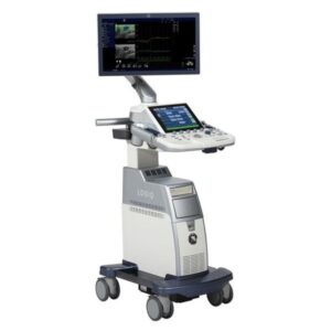
Logiq E10
Ultrasound
B-FlowTM imaging:
B-Flow Capture with Reconstruction: Provides a three- dimensional view of blood vessels
B-Steer+: Enhances needle visualization in real time to improve speed and confidence in needle guidance procedures
Contrast Enhanced Imaging
2D Shear Wave Elastography: enables quantitative assessment of tissue elasticity
Strain Elastography is also available for qualitative tissue assessment.
Volume Navigation: The combination of 2D/3D GPS Tracking and Fusion Imaging
Fusion Imaging: Merge real-time ultrasound with a volume DICOM® MR dataset to facilitate a second look examination and fusion guidance of biopsies of masses seen on MRI but not on ultrasound
2D/3D GPS Tracking
Dual Image CF Pancreas, C1-6-D
Photo Assistant App
Compare Assistant
GE Voluson E6
Ultrasound
40 frames per second in 4D mode
2D/3D/4D Imaging
M-Mode
M-Color Flow
PW/CW Spectral Doppler
Color/Power/Tissue Doppler
HD Flow
B-Flow
GE Voluson E8
Ultrasound
Digital Beamformer
1,979,578 System Processing Channel Technology
Displayed Imaging Depth: 0 – 30 cm
Minimum Depth of Field: 0 – 1 cm (Zoom, probe dependent)
Maximum Depth of Field: 0 – 36 cm (probe dependent)
Up to 274 dB Dynamic Range
GE Voluson E10
Ultrasound
3D/4D imaging
Enhanced resolution and increased frame rates for superior image quality
23 inch widescreen LED monitor
12.1 inch touch panel with multi-touch
Probe port illumination
Fast and secure data management
1-button control panel up/down function
Fast and secure data management
Easy-to-use automation tools to help streamline workflow
Email exports directly from system
GE Vivid E9
Ultrasound
Available in 2D or 4D
Customizable Scan Assist and Scan Assist Pro to quickly acquire images
Multidimensional imaging
Auto-align for 4D views, features automatic positioning and orientation of the left ventricle
Automated imaging options for easy acquisition, enhanced image quality.
Image quality optimization for increased efficiency with a wide range of patients.
Automated speckle-tracking strain and Auto-EF tools.
Numerous connectivity options streamline workflow
GE Venue Point of Care
Ultrasound
Seamless display. For easy cleaning.
ECG and TEE functional.
Backed by a multi-year warranty. Support you can rely on.
Chassis with large wheels and small footprint. Designed for robust use in the emergency room. Simply push the Venue where you need it.
Handy key probe. Control the system with freely programmable keys on the probe handle.
Ready to use when you need it. Up to 4 hours of battery power and a clearly visible remaining time indicator that changes colour as the battery level drops.
Large 19-inch LCD multitouch monitor on height-adjustable swivel arm.
Four probe connections. For a wide variety of examination possibilities.
Intelligent cable management. Four connected and ready to use probes. Well-placed brackets help keep cables away from the ground.
GE Venue 50 Point of
Care Ultrasound
Easy one-step setup for probe and procedure
Fast power-up
Tablet-like navigation
Wireless and portable
Streamlined design for simple operation
Precise imaging
GE Vscan
Ultrasound
Phased Array and Linear transducers in a single probe
Phased Array Transducer for deep scanning:
Field-of-view for black and white imaging: aperture width of 2.9 cm with maximum depth of 8 cm
Imaging Presets including black and white for anatomy and color-coded blood flow
Directional Color Flow, Voice Recorder, Auto Image Optimization
Access to VScan Web Portal / VScan Gateway Software for PC
Storage capacity: 4GB upgradable to 32GB on microSD or microSDHC cards
Data storage formats: .jpg for still frames, .mpg for loops, .wav for voice recordings
Recharge time for device battery: 75 minutes for 90% battery capacity
Philips
Philips iU 22
Ultrasound
B, M, PW Doppler, Colorflow
17” Flat Panel Display
xSTREAM Architecture
PureWave Crystal Transducers
2D Imaging
MPR
iSCAN
iFOCUS
iOPTIMIZE
Panoramic Imaging
Tissue Doppler Imaging
Tissue Harmonics
XRES
SonoCT
Netlink DICOM 3.0
Philips IE 33
Ultrasound
Philips xSTREAM imageformer architecture Up to
442,176 total digital channels (xMATRIX configuration), 57,000 total digital channels (non xMATRIX configuration)
Displayed Imaging Depth: 1 – 39 cm (transducer dependent)
Up to 180 dB system dynamic range
Philips Affiniti
Ultrasound
S5-1 transducer with exclusive Purewave crystal technology provides striking 2D clarity and color sensitivity.
Color-flow mapping: 1
X3-1 with xMatrix array technology incorporates the processing power of 150 computer boards for real-time 3D volume rendered imaging
xMatrix transducer technology incorporates exclusive micro-beamforming circuitry designed to support up to 3,000 individual active elements.
Live xPlane imaging
Philips Epiq
Ultrasound
B, M, PW & CW Doppler, Colorflow
21.5” Flat Panel Display
PureWave Crystal Transducers
TSP
AutoSCAN
Panoramic Imaging
Tissue Doppler Imaging
Tissue Harmonics
AIUS
CEUS
Netlink DICOM 3.0
Philips Epiq CV
Ultrasound
Extraordinary architecture
Superior image quality
xMATRIX is the most leading-edge, versatile ultrasound transducer technology
Q-App quantification applications
nSIGHT Imaging strengthens the power of PureWave to image technically difficult patients
The Mitral Valve Navigator: designed to take a Live 3D volume of the mitral valve and turn it into an easy-to-interpret model in eight guided steps
Portable and quiet
Access to multimodality images
Pro-active monitoring: allows for the detection and repair of anomalies before they become problems
Utilization reports: Data intelligence tools that can help you make informed decisions
Online support request: reduces workflow interruption
Fetal Heart Navigator: allows you to acquire the fetal heart volume in as little as two seconds
iSSL technology: provides privacy standards and a safe and secure connection
Philips CX50
Ultrasound
B, M, PW & CW Doppler, Colorflow
17″ Flat Panel Display
xSTREAM architecture
PureWave Crystal Transducers
2D Imaging
MPR
iSCAN
iFOCUS
iOPTIMIZE
Panoramic Imaging
Tissue Doppler Imaging
Tissue Harmonics
XRES
SonoCT
Netlink DICOM 3.0
Order Today
Reach out to us by clicking below and we will be in touch with you shortly.